Cardiology Diseases – Treatment in Germany
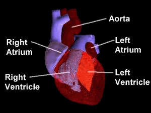
The human heart is primarily a shell. There are four cavities, or open spaces, inside the heart that fill with blood. Two of these cavities are called atria. The other two are called ventricles. The two atria form the curved top of the heart. The ventricles meet at the bottom of the heart to form a pointed base, which points toward the left side of your chest. The left ventricle contracts most forcefully, so you can best feel your heart pumping on the left side of your chest.
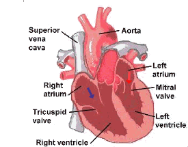
The left side of the heart houses one atrium and one ventricle. The right side of the heart houses the others. A wall, called the septum, separates the right and left sides of the heart. A valve connects each atrium to the ventricle below it. The mitral valve connects the left atrium with the left ventricle. The tricuspid valve connects the right atrium with the right ventricle.
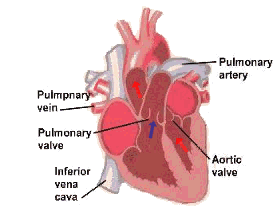
The top of the heart connects to a few large blood vessels. The largest of these is the aorta or main artery, which carries nutrient-rich blood away from the heart. Another important vessel is the pulmonary artery, which connects the heart with the lungs as part of the pulmonary circulation system. The two largest veins that carry blood into the heart are the superior vena cava and the inferior vena cava. They are called “vena cava” because they are the “heart’s veins.” The superior is located near the top of the heart. The inferior is located beneath the superior.
The heart’s structure makes it an efficient, never-ceasing pump. From the moment of development through the moment of death, the heart pumps. The heart, therefore, has to be strong. The average heart’s muscle, called cardiac muscle, contracts and relaxes about 70 to 80 times per minute without you ever having to think about it. As the cardiac muscle contracts it pushes blood through the chambers and into the vessels. Nerves connected to the heart regulate the speed with which the muscle contracts. When you run, your heart pumps more quickly. When you sleep, your heart pumps more slowly.
Considering how much work it has to do, the heart is surprisingly small. The average adult heart is about the size of a clenched fist and weighs about 11 ounces (310 grams). Located in the middle of the chest behind the breastbone, between the lungs, the heart rests in a moistened chamber called the pericardial cavity which is surrounded by the ribcage. The diaphragm, a tough layer of muscle, lies below. As a result, the heart is well protected.
CIRCULATIONCHART:
SUPERIOR & INFERIOR VENACAVA —->RIGHT ATRIUM —->TRICUSPID VALVE —->RIGHT VENTRICLE —->PULMONARY VALVE —->PULMONARY ARTERY —->LUNGS —-> PULMONARY VEIN —->LEFT ATRIUM —->MITRAL VALVE —->LEFT VENTRICLE —->AORTIC VALVE —->AORTA —->TISSUES OF THE BODY.
Internal circulation of the heart is maintained by the network of coronary arteries. Three arteries feed the heart muscle or myocardium. The left anterior descending coronary artery, the left circumflex coronary arteryand the right coronary artery.
The nerve structure of the heart is important as arrythmias and therapies used to treat them frequently invoke these terms. Clusters of nerve cells that give rise to the hearts electrical impulse are called the sinus or SA node, and AV node. The impulse then travels in the likewise manner.
SA node —->Atrial muscle —->AV node —->Common bundle —->Bundle branches —->Purkinje fibers —-> Ventricular muscle. This sequence is represented in an ECG recording.
DISEASES AND TREATMENT
ANGINAPECTORIS
Whatisanginapectoris?
Angina pectoris is the medical term for chest pain due to coronary heart disease. This is a condition in which the heart muscle doesn’t receive enough blood, resulting in pain in the chest.
Angina is a symptom of a condition called myocardial ischemia . It occurs when the heart muscle (myocardium) doesn’t get as much blood (hence as much oxygen) as it needs for a given level of work. Insufficient blood supply is called ischemia.
Whendoesanginapectorisoccur?
Angina pectoris can occur when blood flow to the heart is enough for normal needs but not enough when the heart’s needs increase. It may happen during physical exercise, strong emotions, or extreme temperatures. Running to catch a bus, for example, could trigger an attack of angina while walking to a bus stop might not. Some people, such as those with a coronary artery spasm, may have angina when they’re resting.
ADAMS-STOKES DISEASE
What is Adams-Stokes disease?
Adams-Stokes disease is a transient condition caused by a heart rhythm disorder. It involves fainting, with or without convulsions. In this condition, the normal heartbeat passing from the upper chambers of the heart to the lower chambers is interrupted. This results in a condition called a “heart block”. When a heart block occurs, the heart rate usually slows considerably. This can result in inadequate blood flow to the brain and fainting. This condition is also referred to as Stokes-Adams or Morgangni, Adams-Stokes disease.
ARRHYTHMIAS
What are arrhythmias?
Arrhythmias or dysrhythmias are abnormal rhythms of the heart. Arrhythmias cause the heart to pump less effectively.
Normally the heartbeat starts in the right atrium when a special group of cells (the sinus node or “pacemaker” of the heart) sends an electrical signal. This signal spreads throughout the atria and to the atrioventricular (A-V) node. The A-V node connects to a group of fibers in the ventricles that conduct the electric signal. The impulse travels down these specialized fibers to all parts of the ventricles. This exact route must be followed to ensure the heart pumps properly.
What is a normal heart rate or pulse?
As the electrical impulse moves through the heart, the heart contracts. This normally occurs 60 to 100 times a minute, each contraction represents one heartbeat. The atria contract a fraction of a second before the ventricles. This lets them empty their blood into the ventricles before the ventricles contract.
What causes arrhythmias?
Under some conditions almost all heart tissue is capable of starting a heartbeat. In other words, another part of the heart can become the pacemaker. An arrhythmia occurs:
>> When the heart’s natural pacemaker develops an abnormal rate or rhythm,
>> When the normal conduction pathway is interrupted or
>> When another part of the heart takes over as pacemaker.
What are the symptoms and treatments for slow heartbeating?
These problems can produce a heartbeat that is either too slow or too fast. Excessive slowing of the heartbeat or bradycardia can cause fatigue, dizziness, lightheadedness, fainting or near-fainting spells. These symptoms due to slow heart beating can be easily corrected with an electronic pacemaker that is implanted under the skin.
What are the symptoms and treatments for rapid heartbeating?
Rapid heart beating, called tachycardia or tachyarrhythmia , can produce symptoms of palpitations, rapid heart action, dizziness, lightheadedness, fainting or near fainting if the heart beats too fast to circulate blood effectively. It may be either regular or irregular in rhythm.
When rapid heart beating arises in the ventricles - called ventricular tachycardia – a life-threatening situation can arise. The most serious cardiac rhythm disturbance is called ventricular fibrillation where the lower chambers are quivering and the heart cannot pump any blood. Collapse and sudden death follows unless medical help is provided immediately.
If treated in time, ventricular tachycardia and ventricular fibrillation can be converted into normal rhythm with electrical shock. Rapid heart beating can be controlled with medications by identifying or destroying the focus of rhythm disturbances. These days one effective way of correcting these life-threatening rhythms is by using an electronic device called an implantable cardioverter / defibrillator.
Blood clots can form during atrial fibrillation, a disorder found in close to 2 million Americans. In atrial fibrillation the two small upper chambers of the heart, the atria, quiver instead of beating effectively. Blood isn’t pumped completely out of them when the heart beats, allowing the blood to pool and clot. If a piece of the blood clot in the atria becomes lodged in an artery in the brain, a stroke (or brain attack) results. About 15 percent of strokes occur in people with atrial fibrillation.
ATRIALFIBRILLATION
What is Atrial fibrillation?
Atrial fibrillation is a disorder found in close to 2 million Americans. In it the two small upper chambers of the heart, the atria, quiver instead of beating effectively. Blood isn’t pumped completely out of them when the heart beats, allowing the blood to pool and clot. If a piece of the blood clot in the atria becomes lodged in an artery in the brain, a stroke (or brain attack) results. About 15 percent of strokes occur in people with atrial fibrillation.
How is Atrial fibrillation treated?
Aspirin and warfarin are two medications currently used. They interfere with clotting of blood, thus helping to reduce stroke risk in people with atrial fibrillation. A nationwide study reported in December 1996 found that less than 40 percent of patients with atrial fibrillation were taking warfarin. Anticoagulants are also given to persons who have atrial fibrillation.
Treating atrial fibrillation as an important way to help prevent stroke (or brain attack). For this reason, the American Heart Association recommends aggressive treatment of this heart arrhythmia.
The AHA states the following:
>> A patient who has atrial fibrillation should be treated by his or her physician with some form of preventive medication.
>> Aspirin and warfarin, the currently used medications, can have a major beneficial effect on public health in the United States.
>> Physicians differ on the choice of drugs to prevent embolic stroke ó stroke caused by an embolus (blood clot). It is clear that warfarin is more effective against this type of stroke than aspirin. However, warfarin has side effects, especially in older patients.
>> Warfarin in well-regulated doses that lead to a moderate interference with clotting is effective and appears safe in many patients.
Patients at high risk for stroke should probably be treated with warfarin rather than aspirin unless there are contraindications. Examples include potential bleeding problems or ulcer. Patients over 75 should be followed especially carefully.
AORTICREGURGITATION
What is Aorticregurgitation?
The aortic valve is between the left ventricle and the aorta. Regurgitation means that the valve does not close properly and blood can leak backward through it. The left ventricle has to then pump more blood than normal and will gradually increase in size because of the extra workload. Aortic regurgitation can range from mild to severe. Some people may notice no symptoms at all for years. As the condition worsens, however, symptoms will appear. These can include Fatigue (especially during periods of increased activity), Shortness of breath, Edema (retention of fluid) in certain parts of the body such as the ankles, Heart arrhythmias (abnormal heart beats) or Angina pectoris (chest pain).
What causes Aortic regurgitation?
Aortic regurgitation can be caused by several things. It may be due to a bicuspid aortic valve. This is a congenital (existing at birth) deformity of the valve in which the valve has two cusps (flaps) rather than the normal three cusps. It can also be found in other kinds of congenital heart disease. Aortic regurgitation can also be caused by infections of the heart, such as rheumatic fever or infective endocarditis . Still other causes include diseases that can cause the aortic root (the part of the aorta attached to the ventricle) to widen, such as the Marfan syndrome or hypertension.
ATHEROSCLEROSIS
What is atherosclerosis?
Atherosclerosis is a type of arteriosclerosis. It comes from the Greek words athero (meaning gruel or paste) and sclerosis (hardness). It involves deposits of fatty substances, cholesterol, cellular waste products, calcium and fibrin (a clotting material in the blood) in the inner lining of an artery. The build-up that results is called plaque.
Arteriosclerosis is a general term for the thickening and hardening of arteries. Some hardening of arteries normally occurs when people grow older.
Plaque may partially or totally block the blood’s flow through an artery. Two things that can happen where plaque occurs are:
>> Bleeding (hemorrhage) into the plaque.
>> Formation of a blood clot (thrombus) on the plaque’s surface.
If either of these occurs and blocks the entire artery, a heart attack or Stroke may result.
Atherosclerosis affects large and medium-sized arteries. The type of artery and where the plaque develops varies with each person.
Atherosclerosis is a slow, progressive disease that may start in childhood. In some people this disease progresses rapidly in their third decade. In others it doesn’t become threatening until they’re in their fifties or sixties.
BUNDLEBRANCHBLOCK
What is the normal condition?
In the heart we have a normal pacemaker. This is a specialized group of cells located in the right upper chamber (right atrium) of the heart. Somewhere between 60 and 100 times a minute, this pacemaker emits an electrical impulse. This impulse then travels throughout the heart on a specified route. As the impulse passes through the heart, the heart muscle contracts (or beats). The impulse first travels through the upper chambers (the atria). Before it can go to the lower chambers (the ventricles) it must pass through one small group of cells called the A-V node. The A-V node is located between the atria and the ventricles. After the impulse goes through this A-V node, it goes along a track called the bundle of His (hiss). From there, this bundle divides into a right bundle and the left bundle. These two bundles go to the right and left lower chambers of the heart. All of this is much like following the roads on a freeway map.
What is bundlebranchblock?
Normally, the electrical impulse travels down both the right and left branches at the same speed. Thus, both ventricles contract at the same time. But occasionally, there’s a block in one of the branches. This doesn’t mean that one of the ventricles won’t contract. It just means that impulses must travel to the affected side by a detour and this detour is slower. Therefore, one ventricle contracts a fraction of a second slower than the other side. Usually if there’s nothing else wrong, a person with bundle branch block shows no symptoms. But since we can record the electrical impulses through the heart with an electrocardiogram (E.K.G.), a bundle branch block shows up on the EKG as an abnormality.
If you have bundle branch block, it may have only been noticed when you had an EKG. You may feel fine. However, there’s something wrong with the blocked bundle. For instance, it might mean that a small part of your heart is not receiving enough oxygen-rich blood. Therefore, if you have bundle branch block, your physician will want to see you regularly to be sure no other changes occur. You may have bundle branch block for many years and still feel fine, but it’s important to have regular check-ups.
BACTERIALENDOCARDITIS
Bacterial endocarditis is an infection of the heart valves or the lining of the heart. It occurs when bacteria in the bloodstream (bacteremia) lodge on abnormal heart valves or other damaged heart tissue.
Endocarditis rarely occurs in people with normal hearts. However, people who have certain preexisting heart defects are at risk for developing endocarditis when a bacteremia occurs.
The AHA’s recommendations are especially important for physicians and dentists who care for:
>> Patients with prosthetic heart valves;
>> Patients with a previous history of endocarditis; and
>> Patients with congenital and acquired heart defects such as:
>> Most congenital cardiac malformations
>> Damaged heart valves
>> Hypertrophic cardiomyopathy
CONGESTIVE HEART FAILURE
Congestive heart failure (or heart failure) is a condition in which the heart can’t pump enough blood to meet the needs of the body’s other organs. This can result from:
>> Narrowed arteries that supply blood to the heart muscle – coronary artery disease.
>> Past heart attack, or myocardial infarction , with scar tissue that interferes with the heart muscle’s normal work.
>> High blood pressure.
>> Heart valve disease due to past rheumatic fever or other causes.
>> Primary disease of the heart muscle itself, called cardiomyopathy .
>> Defects in the heart present at birth – congenital heart disease.
>> Infection of the heart valves and/or heart muscle itself – endocarditis and/or myocarditis.
The “failing” heart keeps working but doesn’t work as efficiently as it should. People with heart failure can’t exert themselves because they become short of breath and tired.
As blood flow out of the heart slows, blood returning to the heart through the veins backs up, causing congestion in the tissues. Often swelling (edema) results, most commonly in the legs and ankles, but possibly in other parts of the body as well. Sometimes fluid collects in the lungs and interferes with breathing, causing shortness of breath, especially when a person is lying down.
Heart failure also affects the ability of the kidneys to dispose of sodium and water. The retained water increases the edema.
PREMATURE VENTRICULAR CONTRACTIONS
What are premature ventricular contractions?
Premature ventricular contractions (PVC), also known as “extrasystoles” are “extra” heartbeats arising from an irritable area in the lower pumping chambers or ventricles . They interrupt the normal heart rhythm and cause an irregular beat, often felt as a “missed beat” or a “flip-flop” in the chest. While often benign, when PVC’s occur very frequently or repetitively, they can lead to more serious rhythm disturbances.
What is ventricular bigeminy?
Ventricular bigeminy is one example of a PVC. In this category of irregular or extra heartbeats, a regular heartbeat is coupled with an irregular beat.
ANEURYSM
What is ananeurysm?
An aneurysm is a ballooning-out of the wall of an artery, vein or the heart due to weakening of the wall by disease, injury or an abnormality present at birth. They are often caused or aggravated by high blood pressure. Aneurysms aren’t always life-threatening, but serious consequences such as a Stroke (or brain attack) can result if one bursts in the brain. If an aneurysm on a large blood vessel or the heart wall bursts, a person could bleed to death.
How is ananeurysm detected?
An aneurysm can be detected by x-ray or by imaging techniques such as echocardiography, an (M.R.I.)magnetic resonance imaging or a computed tomography (C.T.) scan. The aneurysm may be small and not cause symptoms. The person’s doctor will want to check it regularly to see if it is enlarging. A common symptom of aneurysm is pain in the area where it is located. The larger the aneurysm becomes, the more likely it is to burst.
How is ananeurysm treated?
Treatment of a brain aneurysm is surgery, during which a metal clip is secured around the base of the aneurysm.
AORTICANEURYSM
What is anaorticaneurysm?
An aneurysm is an area of bulging, much like a bulge on an innertube when it’s overinflated. The aorta, the main blood vessel leading away from the heart, can sometimes develop an aneurysm. The wall of the aorta may have become damaged by atherosclerosis and part of it has become weakened. The condition may also be due to an inherited disease such as the Marfan syndrome. The aneurysm usually occurs in the abdominal area below the kidneys (abdominal aneurysm), but may occur in the chest cavity (thoracic aneurysm). The danger of an aneurysm is that it may burst.
How is ananeurysm detected?
An aneurysm can be detected by x-ray or by imaging techniques such as echocardiography , an M.R.I. (magnetic resonance imaging) or a computed tomography (C.T.) scan. The aneurysm may be small and not cause symptoms. The person’s doctor will want to check it regularly to see if it is enlarging. A common symptom of aneurysm is pain in the area where it is located. The larger the aneurysm becomes, the more likely it is to burst.
How is ananeurysm treated?
Treatment is surgery, during which a patch or artificial piece of blood vessel is sewn in place where the aneurysm was.
DIASTOLICDYSFUNCTION
What is diastolicdysfunction?
The heart contracts and relaxes with each heartbeat. The contraction portion of this cycle is called systole and the relaxation portion is called diastole. If the relaxation part of the cycle is abnormal, this is referred to as diastolic dysfunction. In some people with heart failure, the contraction function is normal but there is impaired relaxation of the heart (specifically, the ventricles) . Because the ventricle doesn’t relax normally, the pressure in it increases to a larger than normal value as blood for the next heartbeat enters it (it is harder for all of the blood to go into the ventricle). This can cause increased pressure and fluid in the blood vessels of the lungs – pulmonary congestion or increased pressure and fluid in the blood vessels coming back to the heart – systemic congestion. People with certain types of cardiomyopathy may also have diastolic dysfunction.
HEARTMURMURS
What causes heartmurmurs?
Heart murmurs can be caused by defective heart valves or holes in the heart walls. A stenotic heart valve cannot open completely, so blood is ejected through a smaller than normal opening. A valve may also be unable to close completely, leading to regurgitation of blood back through the valve when it should be closed – such as aortic or mitral regurgitation. Murmurs can also be caused by conditions such as pregnancy, fever, thyrotoxicosis or anemia.
A diastolic murmur occurs during heart muscle relaxation between beats. A systolic murmur occurs during heart muscle contraction. Systolic murmurs are graded by intensity (loudness) from one to six. For example, a grade 1/6 is very faint, heard only with a special effort. A grade 6/6 is extremely loud and heard with stethoscope slightly removed from the chest.
LONGQ-TSYNDROME
What is the longQ-Tsyndrome(LQTS)?
Long Q-T syndrome (L.Q.T.S.) is an infrequent, hereditary disorder of the heart’s electrical rhythm that can occur in otherwise healthy people. It usually affects children or young adults. It is often inherited and is usually congenital (present from birth).
When the heart contracts, it emits an electrical signal. This signal can be recorded on an electrocardiogram (E.K.G. or E.C.G.) and produces a characteristic waveform. The different parts of this waveform are designated by letters – P, Q, R, S and T. The Q-T interval represents the time for electrical activation and inactivation of the ventricles , the lower chambers of the heart. A doctor can measure the time it takes for the Q-T interval to occur (in fractions of a second), and can tell if it occurs in a normal amount of time. If it takes longer than normal, it is called a prolonged Q-T interval.
MITRALVALVEANDMITRALVALVEPROLAPSE
The mitral valve is the heart valve between the left atrium and left ventricle . It has two flaps or cusps.
What is mitralvalveprolapse?
In mitral valve prolapse (M.V.P.), one or both valve flaps are enlarged and some of their supporting “strings”may be too long. When the heart contracts or pumps, the mitral valve flaps do not close smoothly or evenly. Instead, part of one or both flaps collapses backward into the left atrium. This sometimes allows a small amount of blood to leak backward through the valve and may cause a heart murmur.
PERICARDIUMANDPERICARDITIS
What is the pericardium?
The pericardium is the thin sac (membrane) that surrounds the heart and the roots of the great vessels.
What is pericarditis?
Pericarditis is inflammation of the pericardium. The pericardium has an inner and outer layer and contains a small amount of lubricating fluid between the layers. When the pericardium becomes inflamed, the amount of fluid between its two layers increases, compressing the heart and restricting its action.
RAYNAUD’S PHENOMENON
What is Raynaud’s phenomenon?
Raynaud’s phenomenon is a condition in which the smallest arteries that bring blood to the fingers or toes constrict (go into spasm) when exposed to cold or as the result of emotional upset. Working with vibrating machinery or smoking cigarettes also can bring on these episodes. The small veins are usually open, so the blood drains out of the capillaries , and the fingers or toes become pale, cold and numb. If there’s a spasm in the small veins and blood is trapped in the capillaries, the fingers or toes turn blue as the blood loses its oxygen.
Raynaud’s phenomenon is also sometimes called Raynaud’s syndrome or Raynaud’s disease.
RHEUMATICHEARTDISEASE/RHEUMATICFEVER
What are rheumaticheartdisease and rheumaticfever?
Rheumatic heart disease is a condition in which the heart valves are damaged by a disease process that begins with a strep throat from streptococcal infection. If it’s not treated, the streptococcal infection can develop into acute rheumatic fever.
Rheumatic fever is an inflammatory disease that can affect many connective tissues of the body – especially those of the heart, joints, brain or skin. Permanent heart damage from rheumatic fever is called rheumatic heart disease.
Anyone can get acute rheumatic fever, but it usually occurs in children five to 15 years old. The resulting rheumatic heart disease can last for life.
What are the symptoms of rheumaticheartdisease?
The symptoms vary greatly from person to person. Often the damage to heart valves isn’t immediately noticeable. A damaged heart valve either doesn’t completely close or doesn’t completely open.
Eventually damaged heart valves can cause serious, even disabling, problems. These problems depend on the severity of the damage and on which heart valve is affected. The most advanced condition is congestive heart failure.
PERIPHERALVASCULARDISEASE
What is peripheral vascular cardiology disease?
Peripheral vascular disease refers to diseases of any of the blood vessels outside of the heart and to diseases of the lymph vessels. It is often a narrowing of the blood vessels that carry blood to leg and arm muscles. There are two types of these circulation disorders:
Functional peripheral vascular cardiology diseases are not organic in cause and do not involve defects in the structure of the blood vessels. They are short-term effects and may be reversed. An example is Raynaud’s disease (or Raynaud’s phenomenon). It can be triggered by cold temperatures, emotional stress, work with vibrating machinery or smoking.
Organic peripheral vascular cardiology diseases are caused by structural changes in the blood vessels (such inflammation and tissue damage). An example is Buerger’s disease.