UPPEREXTREMITY
The upperextremity generally comprises of shoulder, elbow, forearm, and wrist.
Pronation is the act of rotating the forearm so the palm is turned backward or downward.Supination is the act of rotating the forearm so the palm isturned forward or upward. The muscle that flex the wrist andfingers are bunched together along the volar surface of theforearm and are attached by a common tendon to the medialepicondyle of the humerus.
WRIST
The wrist is made up of the distalends of the radius and ulna plus eight carpal bonesarranged in a proximal and distal row of bones. Stability of thewrist is maintained by strong ligaments that lash the carpalbones together. The hand is made up of five metacarpal bones and the phalanges of four fingers and the thumb.
LOWEREXTREMITY
ANATOMYOFTHEHIPJOINT
The lower extremities carry the bodyweight and must provide for locomotion, the joints of this partof the body are big, tend to be strong and stable, and are served by strong muscles.
The hip joint is a close-fitting, deeplyset ball and socket joint between the rounded head of the femur (ball) and the acetabulum (socket) on the side of the pelvic iliacbone and is further supported by a surrounding strong capsule.The principal hip flexor muscle, the iliopsoas, is located infront of the joint. Posteriorly, the gluteus maximus musclefunctions as the principal hip extensor. The gluteus maximus andmedius muscles abduct the thigh and the adductor muscles(adductor longus, brevis, and magnus, and the gracilis muscles),grouped together along the inner surface of the thigh, adduct the thigh.


BASICANATOMYOFTHEKNEEJOINT
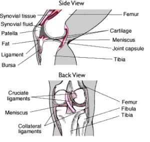
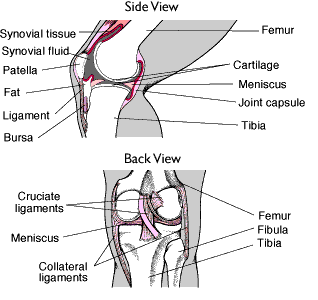
The knee, the largest joint in the body is a hinge joint between the femur and the tibia that permits the motions of flexion and extension. In common with other axial joints, the knee joint is lined with a thick synovial membrane that secretes synovial fluid to lubricate the joint surfaces. With an injury or inflammation of the knee joint this membrane often produces a large amount of fluid that often causes pain for the individual.
ANATOMYOFTHEFOOT
The human foot consists of 26 bones, 33joints, 11 muscles, and a myriad of nerves, arteries, veins, and lymphatic channels.
Whatarethedifferentpartsoffootbone?
The foot bones are talus (ankle bone),calcaneus or os calcis (heel bone), cuboid, cuneiform bones (three in number), navicular, metatarsals (five in number),phalanges (two for the hallux, or great toe; three for each ofthe other digits).
Other bones include two sesamoid bones beneath the first metatarsophalangeal joint ( a medial andlateral sesamoid bone, also known as a tibial or fibular sesamoidbone). Additional bones infrequently seen in the human foot arethe os navicularis (an extra bone medial to the navicular bone),the os trigonum (behind the talus), and the os peroneus (lateralto the cuboid bone).
Collectively, bones in the toes areknown as phalanges, and bones proximal to this are known asmetatarsals. Bones in the mid-foot to the rear foot are known astarsal bones. The talus, or ankle bone, articulates with the two lower leg bones — on the lateral side or the outer side as the fibula and the inner border as the tibia. The distal end of thesebones are known as the lateral malleolus for the fibula and medial malleolus for the tibia.
The top of the foot contains only oneintrinsic muscle, known as the extensor digitorium brevis muscle.It sends tendons to the great toe, or hallux, and to the next three toes. This muscle is known as brevis because it inserts atthe more proximal aspect of the toes in its job of extending orbringing up to the toes. Extrinsic muscles that arise outside thefoot end as tendons and insert into the distal phalanges. Theyhave the same function of extension and are known as the extensordigitorium longus muscle, which arises in the front of the lowerleg.
DISEASESANDTREATMENTSOFFOOTDISEASES
INTEGUMENTARYDISEASES
CORNS:
HARD CORNS are also known as heloma durum and are usually found on top of the toe.
SOFT CORNS are also known heloma mole and are most frequently found between the small and fourth toes.
CALLUS are compacted tissue lesions on the bottom of the foot beneath the metatarsal bones; they are often referred to as intractable plantar keratoma (IPK).
WARTS are viral infections of the skin. If they are on top of the foot they are known as verruca vulgaris. Warts on the bottom of the foot do not extend outwards but are embedded within the foot are known as Veruca Plantaris or plantar wart.
FOLLICULITIS is the infected hair follicles.
ONYCHOMYCOSIS is the fungal infections of the toe nail
ONYCHOCRYPTOTIC is ingrowing toe nail.
PSORIASIS is the systemic skin disease that can be manifested in the foot and nails.
PARONYCHIA is an ingrown toe nail.
ONYCHOCHAUXIS is the simple thickening of the toe nail without evidence of infection.
ONYCHOLYTIC is a toe nail loosened from the nail bed.
TINEA PEDIS is the fungal infection of the feet.
CELLULITIS is the inflammation of the soft tissue.
PERIPHERALVASCULARDISEASESDIABETICFOOTDISEASE:
Diabetes involves thickening of the basement membrane of the capillary, and this ultimately leads to malnourishment of peripheral nerves, leading to a progressive and inevitable diminished sensation in the extremities known as diabetic neuropathy. When there is extensive calcification usually associated with diabetes, the condition is known asarteriosclerosis obliterans.
YMPHATICDISEASES:
These are manifested by edema, or fluid collection in soft tissue. When a thumb is pushed into the swollen area, a deep impression is made, leaving a pit, it is known aspitting edema.
VARICOSITIES:
When the delicate one way valves of veins become compromised and allow leakage of fluid backward, creating more back pressure on the veins farthest from the heart, it is associated with a darkening of skin about the malleoli, known as varicosities or varicose veins.
ULCERATIONS:
These are compromises in the skin. These ulcerations, if associated with the veins, are around the ankle, and if they are associated with the arterial supply or with diabetes due to neuropathy, they are on the plantar aspect of the foot.
NEUROLOGICDISEASES
MUSCLEWASTINGANDATROPHY:
The sensory and motor nerves are seen in pathologic conditions with diminished ability to create appropriate sensation signals that are sent to the brain. This is associated with diabetes, alcoholic neuropathy, leprosy, and other diseases.
MUSCULOSKELETALDISEASES
Whatisatypicalpartofmusculoskeletalexamination
The musculoskeletal examination is very specific, and detailed because the mechanism of the injury is a very important part of the initial history. This is because the position of the foot or ankle will predict type and severity of injury regardless of what physical signs or symptoms show. The circumference of the calf muscle is measured, range of motion of all the peripheral joints is examined in detail, and compared with the normal values ( the normal values can be obtained from the side that is not injured). The gait analysis and stance examination is a typical part of any biomechanical or musculoskeletal examination. A bunion is the prominence on the inner border of the foot at the first metatarsal head. The lateral drifting of the big toe or the hallux often associated with this is known a hallux valgus.
After examination of the gait and stance, the foot is examined for the presence of digital deformities aside from hallux valgus. A common digital deformity is hammertoe, which is a contracture of the proximal interphalangeal (PIP) joint, leaving the first bone or proximal phalanx dorsi flexed onto the metatarsal bone behind it, with the middle and distal phalanges being flexed. This is associated with the formation of a hard corn at the dorsal aspect of the proximal interphalangeal joint.
Another digital deformity is a mallet toe, a contracture of the end joint or the distal interphalangeal (DIP) joint, which is the end bone being plantar flexed towards the ground. This is associated with a hard corn on the dorsal aspect of the DIP joint.
Inflammation of the metatarsophalangeal (MP) joints is called capsulitis, or inflammation of the capsule. This can be affiliated with a periarticular bursitis or bursa formation and is sometimes associated with or found to be a cause of neuritis, an inflammation of the nerve that runs between the metatarsophalangeal joints. The most common is Morton’s neuroma, found between the third and fourth metatarsal heads.
The presence of heel spur or a projection from the bottom of the heel bone pointing forward is associated with pain at the plantar aspect of the heel with stiffness or inability to walk early in the morning
FRACTURES
Whatarethedifferentfractures?
SIMPLE FRACTURE in which two fragments are created, one proximal and one distal.
COMMINUTED FRACTURE when there are more than two fragments.
COMPOUND FRACTURE in which there is a break of skin by the fractured bony pieces.
COMPRESSION FRACTURE which is a compressing or crushing type of fracture.
STRESS FRACTURE which is resulting from force on a bony structure.
AVULSION OR CHIP FRACTURE when a tendon or ligament pulls a piece of bone away from larger bone.
GREENSTICK FRACTURE seen in children when one cortex is broken and the other bows but does not give.
TREATMENTS
NONSURGICALPROCEDURES
Use of digital pads for treatment of a heloma durum and felt padding or a lamb’s wool pad applied to or around the toe to alleviate pressure from the contracted joint. The soft corn is treated with packing of the interspace with lamb’s wool to separate the proximal phalanges of the two digits involved.
IPK is treated by trimming the lesion and putting a pad around it or by fabrication of an orthotic device. Orthotics are custom molded inserts taken from a cast of the patient’s foot. They are of two types : accommodative orthotics, biomechanical or functional orthotics.
Chronic ulcerations are sometimes treated with an anterior heel, rocker bar, or metatarsal bar.
Dressings of gauze impregnated with zinc oxide paste, known as Unna boot, medico paste bandage, or soft cast is used to treat venous stasis and ulcerations.
In nonsurgical treatment of hallux valgus, or bunion deformity, a molded shoe known as a space shoe, custom made by a shoemaker, is prescribed and fitted. This is done from a plaster casting made of the foot. Use of foam sleeve and a latex molded shield is used to support or protect the bunion. Various other functional orthotic devices are used to treat the bunion deformity.

